Intraoral Lesions
In general, the important issue regarding skin or mucosal lesions inside the mouth is that any sign such as ulcers, discoloration, shape changes, or tissue alterations in the skin or mucous membrane of the mouth, should be examined and given attention if the symptoms persist for more than two weeks. If necessary, a sample should be taken.


An Introduction to Intraoral Lesion Surgery
Upon noticing the first sign in the soft tissue, the patient should visit an oral and maxillofacial surgeon for consultation. After assessing the patient’s clinical condition, the surgeon usually recommends waiting for about two weeks from the onset of the initial symptom. During this time, the patient should follow the hygiene and medication instructions provided by the surgeon. If the condition naturally improves and no further intervention is required, there is no need for additional measures. However, if the condition does not improve after two weeks and the symptoms persist, the oral and maxillofacial surgeon may order radiography, CT scan, or MRI (if necessary) for a more detailed examination and proceed with sample collection to determine the nature of the lesion.
In some cases, your symptoms may resolve after two weeks, but the same symptom may reoccur in the same area over time, in the following months or years. In such cases, after reevaluation and obtaining new radiography or MRI, we proceed directly to sample collection and perform a new biopsy of the area of interest to investigate the cause of the recurring lesion and its nature.
The Surgical Procedure for Intraoral Lesions
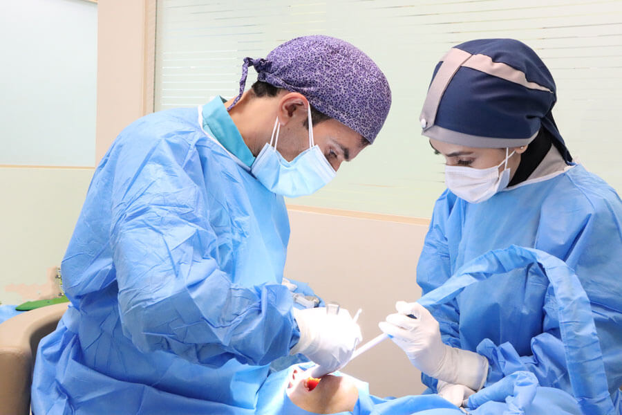
During sample collection, the oral and maxillofacial surgeon will either remove the entire lesion and surrounding tissue or take only a portion of the lesion for pathology. Then, based on the pathology results and the diagnosis of the lesion, they determine the treatment plan and the type of surgery. This varies depending on the size, location, and type of lesion in different individuals and conditions.
An important point to note regarding the removal of the lesion or sample collection and biopsy is that it must be performed by a specialist. This is because a thorough examination of radiography, CT scan, or MRI prior to biopsy, providing the appropriate treatment plan, preserving the tissue at the site of lesion removal during sample collection, and paying attention to lymph node involvement before and during biopsy are all crucial.

Surgical Excision of Benign Lesions
If you have been dealing with an intraoral or facial and neck lesion and have not experienced improvement even after a two-week period, you can reach out to us for consultation with an oral and maxillofacial surgeon.
- Saturdays: 14:00 to 20:00 - Punak clinic
- Sundays: 14:00 to 20:00 - Shariati clinic
- Mondays: 14:00 to 20:00 - Punak clinic
- Thursday: 14:00 to 20:00 - Saveh clinic
Benign Tumors of the Jaw and Face
Benign cysts and tumors of the jaw are growths that develop in the jawbone or the soft tissues of the mouth, jaw, and face. These lesions, also known as odontogenic tumors, can vary in size and rate of growth. At times, they can cause bone destruction and even result in the displacement of teeth. Certain tumors such as ameloblastoma and odontogenic myxoma exhibit an invasive local behavior and require a more extensive form of treatment.
Other common benign tumors affecting the mouth, jaw, and face include odontogenic keratocyst (OKC), central giant cell granuloma (CGCG), radicular cyst, dentigerous cyst, and odontoma.
Upon a definitive diagnosis, a sample of the tumor is obtained. The treatment typically involves the surgical excision of the lesion. In some cases, it may also be necessary to remove adjacent tissues and teeth. Once the tumor is removed, it is sent to the laboratory for further examination.
In certain instances, concurrent or secondary reconstruction of the surgical site is performed, guided by the type of tumor, surgical approach, and the recommendations of the oral and maxillofacial surgeon. The human body itself often compensates for the lost tissue, which depends on the patient’s physical condition and the characteristics of the tumor. Postoperative medication and regular follow-up visits to the physician are imperative components of the overall treatment.

Biopsy
Biopsy involves the surgical removal of a sample from the site of the lesion for examination, determination, and diagnosis of the nature of the lesion, be it malignant or benign.
The excised tissue is sent to a specialized oral and maxillofacial pathology department, where a pathologist analyzes the sample using microscopic examination or chemical agents. This procedure demands a high level of precision and sensitivity, yet in terms of the treatment process, it has a short surgical time.
Anesthesia is often unnecessary, and the procedure is typically performed under local anesthesia, similar to tooth extraction. However, certain cases may necessitate anesthesia if access to the lesion is challenging, there is a significant risk of bleeding, or if the patient has uncontrolled underlying medical conditions.
Through a small incision made inside the oral cavity, the surgeon exposes the soft tissue and gains access to the site of the lesion. The lesion is then excised for subsequent analysis by the pathology department, and the surgical site is briefly sutured.
Soft tissue lesions that may require biopsy include:
- Recurrent ulcers such as leukoplakia and erythroplakia
- Ulcers or lesions that persist for more than two weeks or demonstrate progressive growth into adjacent areas
- Lesions causing discoloration of the affected region or surrounding skin
- Lesions that continue to grow despite regular check-ups and evaluations
- Lesions presenting noticeable signs, such as discharge, infection, pain, inflammation, etc
- Lesions that are initially controlled with medication and hygiene instructions but subsequently recur over time


Hard tissue lesions that may require biopsy include:
Lesions that are not visually detectable or palpable, yet present symptoms such as pain, discharge, and inflammation.
Lesions that are observed in radiographs without apparent signs, but it is advisable to remove them based on their location and the potential involvement of adjacent areas.
Lesions originating at the apex of tooth roots that remain unresolved despite root treatment, leading to recurrence or root resorption.
Examples of such hard tissue or intraosseous lesions include tori, exostoses, periapical lesions, radicular cysts, and periapical granulomas
Soft Tissue Irritative Lesions in the Oral Cavity
A subset within the realm of soft tissue lesions in the oral cavity comprises irritative lesions. These lesions arise as a consequence of an inciting factor, and the initial step involves addressing the underlying irritants. The following are instances of irritative lesions:
For instance, an individual experiencing a fractured tooth may inadvertently traumatize the tongue or oral mucosa, resulting in the formation of a painful ulcer. This ulcer may exhibit white or red discoloration and be accompanied by pain, swelling, and inflammation. In this scenario, the sharp edges and fracture of the tooth act as irritants to the tongue or lip, giving rise to the development of an ulcer or lesion.
[read more]
Patients utilizing dentures may encounter bone resorption over time, leading to compromised stability and fit. Excessive ridges on the denture may cause chronic irritation and stimulate the vestibular area, ultimately resulting in the development of lesions such as epulis fissuratum.
In such cases, it is imperative to abstain from using the denture for a period of two weeks to assess whether the lesion diminishes in size or persists. If the lesion does not reduce in size or remnants remain, a decision is made regarding the necessity of surgical intervention and biopsy. In this context, the use of dentures serves as an irritant contributing to the development of the lesion.
In individuals afflicted with dental caries, the carious lesion itself can act as an irritant. Caries can give rise to various lesions such as giant cell granulomas, periapical giant cell granulomas, pyogenic granulomas, and a like. Thus, dental caries can also serve as an inciting factor for irritative lesions.
Improperly fabricated dental restorations or those with excessive margins can provoke irritation of the gingiva, eliciting symptoms in the surrounding soft tissue. These symptoms may encompass excess tissue growth, pain, redness, and bleeding. Morphologically, these lesions are often likened to cauliflower-like in appearance. They typically exhibit a pedunculated nature.
These aforementioned irritative lesions are generally managed by addressing the underlying irritant, and specific treatment is not typically required. However, if the lesions persist or recur, it is imperative to consult with a surgeon for further evaluation and determine the necessity of surgical excision of the lesion.
[/read]

Types of Soft Tissue Lesions in the Oral Cavity
This condition typically arises from the sudden exposure of the soft tissues within the oral cavity such as the lips, labial mucosa, tongue, palate, throat, and tonsils to hot substances during ingestion. It induces burns, swelling, blistering, pain, and associated symptoms. However, with proper care and the removal of the inciting agent, these manifestations are typically alleviated within a two-week period.
Fistulas can manifest in cases where dental root problems, infections within the root canal, intraosseous lesions surrounding or at the apex of the tooth root, or deep periodontal pockets are present. Consequently, the affected gum becomes edematous, giving rise to a soft tissue swelling encapsulating fluid. This is known as a fistula.
Primarily resulting from repetitive erroneous habits or, occasionally, as a consequence of sharp or prolonged tooth biting within a particular region of the cheek. Hence, labial gas entrapment engenders mucosal edema, pain, and the formation of a prominent linear elevation on the adjacent soft tissue surface in close proximity to the tooth. In such cases, the condition can be controlled through behavioral, dietary, and corrective measures, including addressing any improper dental restorations.
Mucoceles arise as a consequence of lip biting and belong to a group of obstructive lesions originating from minor or accessory salivary glands. When the patient bites the minor or accessory salivary glands, salivary gland ducts get obstructed, resulting in the accumulation of saliva beneath the mucosa of the lip. Mucoceles frequently exhibit a bluish hue within the mucosal tissues, with a higher incidence observed in the submucosa of the lower lip. While many cases spontaneously resolve, persistent mucoceles necessitate surgical excision, often accompanied by the removal of accessory glands during the procedure, if required.
This lesion presents as a localized proliferation of tissue within the mucosa, closely resembling excess fleshy growth. Typically arising from biting, irritation fibromas can manifest in the lip, cheek, tongue mucosa, etc. These lesions are usually not worrisome but should be monitored if they persist beyond two weeks. Surgical removal is performed, and the excised tissue is sent for pathology to determine the nature and characteristics of the lesion.
In this condition, the superficial layer of the oral mucosa undergoes destruction.

Post-irritative Lesion Care
- Typically, it is necessary to allow a two-week period for the recovery of the affected soft tissue area.
- During this time, the irritant agent should be removed.
- You may use painkillers if there is pain.
- The use of chlorhexidine mouthwash is recommended twice a day for one week.
- Compliance with hygiene and dietary practices is necessary.
- If the lesion is not resolved or becomes larger after two weeks, surgical intervention is performed to remove the lesion. A sample is sent to pathology for examination and diagnosis of the type and nature of the lesion.
Types of Mucosal Lesions
- Ulcers
- Superficial Mucosal Lesions
- Salivary Gland Lesions
- Salivary Gland Stones
- Ranula
Various factors contribute to the development of ulcers such as medication use, OCLs, smoking, vigorous tooth brushing, dental procedures, etc. Ulcers are observed in different sizes. They are 1 to 2 mm in size and can occur in various locations within the oral soft tissue. They cause pain and are more commonly found in the depth of the vestibule. In certain conditions, such as AIDS where the immune system is severely compromised, the size of ulcers may be larger and more persistent in the oral mucosa. Research indicates that stress is an important predisposing factor for ulceration. Stress leads to the formation of ulcers in the oral mucosa, and the healing period is approximately 21 days. It is preferable to endure any pain or discomfort during this period and avoid manipulating the affected area, allowing it to heal naturally. Ulcers generally do not pose serious problems for the patient, and the inflammation of the area gradually decreases in two weeks. Pain killers can be used if needed. The use of mouthwash also aids during this period. Consult with your physician regarding the type of mouthwash to be used.
A wide range of benign soft tissue lesions can occur in the oral mucosa, each with its own origin. For example, nerve tissue lesions such as Schwannomas have a neural origin and are highly sensitive to trauma and pressure.
Leukoplakia and erythroplakia also cause superficial mucosal irritation. In such cases, proper diagnosis and sample collection are necessary to determine the nature of the lesion through pathology examination.
Some leukoplakia and erythroplakia lesions are precancerous. That is, they have the potential to transform into malignant lesions. Therefore, special attention should be given to these conditions. These lesions are generally large, painful, red, white, and extensive in nature.
There are three main major salivary glands in the mouth: parotid, submandibular, and sublingual. These glands are bilateral and primarily responsible for saliva production. The parotid gland is located in front of the ear, the submandibular gland is located beneath the jaw, and the sublingual gland is located beneath the tongue. All of these glands empty into the oral cavity through a series of ducts.
This is a condition that usually presents itself with pain, swelling, and infection. It is anatomically manifested in the submandibular gland. Sometimes the stone forms within the duct, while other times it forms within the gland itself. One of the main signs of this condition is the patient’s reaction to sour foods. If they experience swelling and enlargement of the lesion after seeing or consuming sour foods, it indicates the presence of a stone. The lesion then shrinks gradually. This sign is observed in the early stages of the disease. When the salivary glands secrete saliva, if a stone obstructs the duct and prevents the saliva from being expelled, it causes accumulation of saliva and enlargement of the duct, leading to subsequent infection. Stones are initially small and non-calcified, so there is a possibility of their expulsion in the early stages. However, if they are not expelled, they grow larger and calcify, leading to the development of infection. In such cases, the first step in diagnosis and treatment is to obtain an orthopantomogram (OPG) or a CT scan and sialography to accurately examine the lesion and its location in three dimensions. The treatment plan is then based on these findings, emphasizing the importance of accurate diagnosis in this condition. Occasionally, this condition occurs in individuals who are physiologically prone to stone formation in the body, such as those with a history of kidney stones, bladder stones, or gallstones. Even if these individuals do not have such a history and only experience salivary gland stones, there is a likelihood of the stones occurring in their body in the future. From a therapeutic perspective, if the salivary gland stone is small, the patient is given hygiene instructions and dietary recommendations and is placed under observation to determine if expulsion is possible. However, if the stone is large, after examining the radiographs and sialography to determine the location of the stone, it is removed through a local surgical procedure. The duct is then cleared and rinsed in the floor of the mouth, and the treatment is carried out. Sometimes, the stone is observed within the gland itself, which makes it impossible to perform the procedure or surgery from inside the mouth. In such cases, the treatment must be carried out externally under anesthesia, and usually, the main gland and duct are also removed. One common side effect of this surgery is a decrease in saliva production, but it does not have a significant impact on the dryness of the mouth.
Swelling and infection of the salivary glands, which usually occur in the parotid gland, also represent a type of salivary gland lesion. This condition is sometimes treated with saliva stimulation and antibiotic therapy.
One of the benign lesions of the salivary glands is observed in the sublingual gland, which is called ranula and is located in the floor of the mouth. This lesion is a salivary retention cyst.
Swelling and infection of the salivary glands usually occur in parotid glands. This is also a type of salivary gland lesion that is sometimes treated with salivary secretion stimulation and antibiotic therapy.
- Plunging Ranula (PR)
It is a cyst-like lesion that extends towards the neck and requires examination, diagnosis, and surgical removal. It should be sent to pathology for further evaluation. One of the diagnostic methods is aspiration in the area of interest to determine if saliva is present inside it. If so, surgery is performed to remove the lesion.
- Plurimorphic Adenoma
This lesion usually has the highest prevalence in the parotid gland and is diagnosed by closed biopsy to determine the appropriate surgical approach based on the surgeon’s opinion and the condition of the lesion. Preservation of the facial nerve branches is of great importance. The parotidectomy being superficial or deep, it needs to be analyzed. The surgical site is accessed through the front of the ear while preserving the facial nerve branches, the lesion is removed, and it is sent to pathology for examination of its type and nature.
An important point to note in all benign soft tissue lesions is that they must be thoroughly examined and evaluated because otherwise, there is a potential for malignant transformation.
Treatment of Salivary Gland Stones
Sialography: Sialography is one of the diagnostic methods for detecting salivary gland stones and determining their location. It is performed by injecting a radiopaque contrast agent and the location of the lesion is determined based on the results, usually by specialized maxillofacial radiologists.
If an oral and maxillofacial surgeon or head and neck cancer specialist determines the need for removal and surgery after the necessary assessments, the lesion is usually removed surgically, and it is sent to pathology for analysis of its type and nature.
Methods such as lithotripsy or other stone-breaking techniques are not very effective or common in salivary gland stones.
Antibiotics are usually prescribed due to the presence of infection.
Hygiene instructions, dietary recommendations, and the use of mouthwashes are provided.
